Common eye conditions
A cataract occurs when the lens inside your eye begins to cloud over. This affects the quality of light getting into your eye and slowly reduces vision. This is generally a slow process but certain types of cataract can change more quickly. Surgery to remove the cataract and implant a plastic lens is indicated when vision is poor enough. Bad vision due to cataract can precipitate falls.
This is a disease that affects the central part of your eye – the macula. The macula is what gives you clear vision. Over time but also due to other factors, this area can be subject to wear and tear effectively. Central vision slowly deteriorates over the course of many years in the ‘Dry’ type. ‘Wet’ AMD can affect vision much more quickly, when fluid leaks into an otherwise fluid-free macula. This can be treated with injections in the hope of making vision better.
Glaucoma is a group of diseases of the optic nerve. The optic nerve consists of hundreds of thousands of nerves leaving the eye to the visual cortex at the back of the brain. It is here that vision is processed. When the nerves die due to Glaucoma, the signal getting through deteriorates and vision begins to get worse. This tends to start in the peripheral area of your vision and is not normally apparent. A visual field test is utilised to pick up early changes. Increased pressure in the eye is the biggest risk factor for Glaucoma. This is why it is always tested at your annual eye test usually starting at 40 years old.
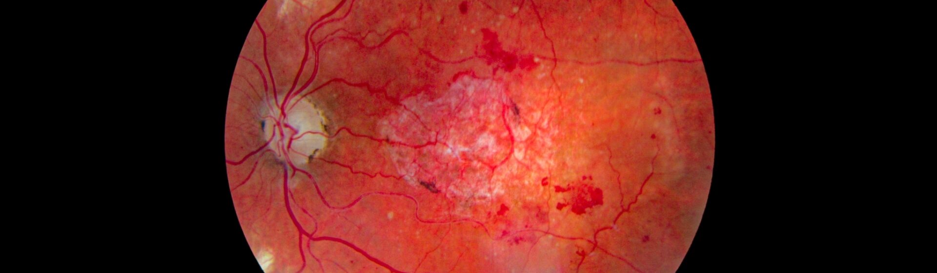
Diabetes often comes with changes to the back of the eye on the retina. This tends to be small haemorrhages and microaneurysms due to the poor calibre of the blood vessels in the eye. This can also lead to hypoxia (lack of oxygen) in the retina causing other features of Diabetic Eye Disease in the retina, including leakage of lipids, disruption in the flow in the nerves leaving the eyes and new blood vessels of poor structure being formed. Diabetes is the greatest cause of working age blindness in the UK. There is a retinal screening service in Scotland that photographs and grades the eyes to assess the risk to sight. This is done on annual basis normally and is instrumental in reducing blindness. Control of Diabetes is of the utmost importance in keeping the eyes as healthy as possible.
This is latin for ‘fleeting loss’. This is when vision is lost in one eye, with everything going black or grey. This generally only lasts for a number of seconds and is caused by a blockage in an artery in the eye, effectively a stroke in the eye. The blockage is released however and vision returns. This is an emergency, as the blockage could then occur in the brain causing a stroke. Visual assessment and referral to the Stroke Clinic is advised immediately.
This is when the retina comes off at the back of the eye. It can cause you to lose vision in the most serious of cases, if the macula comes off as well. This is usually precipitated by flashing lights and floaters in the vision. A grey patch can suddenly appear in your vision or it can feel like a curtain is coming down over your vision. This is an emergency and normally needs surgery to put the retina back onto the back of the eye. Leaving this too long without investigation can mean that vision can be irreversibly damaged, making it a life changing event.
This normally occurs out of the blue in one eye. It occurs in 50% of people over 50 years old. The jelly-like substance that occupies the space in the centre of your eye (vitreous humour) liquifies over time and shrinks. This process starts in your 20’s. Eventually it peels off the retina at the back of your eye, as a perfectly normal event. It can start with flashing lights and floaters. A large floater can appear on the outer side of your vision in either eye, moving as your eye moves. This can present similarly to a retinal detachment, meaning that has to be ruled out. A PVD can take a little while to fully detach, causing flashes to occur intermittently. Occasionally, the vitreous can stick a little more strongly to a part of the retina causing a tear. This in turn can lead to a retinal detachment. It is important to have this checked out urgently.
This is an infection of the lashes and eyelids in your eyes. There are generally two types – posterior and anterior. Symptoms include sticky lashes, debris in the lashes, redness of the lid margin, grittiness and itchy uncomfortable eyes stuck together in the morning. Treatment is the same for both with heating and cleaning of the eyelids. A treatment of around 2 weeks can fix this problem well but sometimes ongoing treatment is required on a less regular basis, as it tends to recur.
The most common causes of this are of bacterial, viral and allergic origins. The tissue on the inside of the eyelids becomes inflamed and red. The white part of the eye can become injected also, with symptoms of burning, grittiness and itching. Viral conjunctivitis tends to just need lubrication for comfort and time for it to resolve. It is very contagious. Bacterial conjunctivitis is also commonly self limiting. Comfort drops and lid cleansing can resolve this but sometimes an anti-biotic is needed to clear it up. Drops for allergic conjunctivitis are aimed at helping the itching and giving comfort to the eye. Anti-allergy drops are commonly needed daily throughout the allergic season. This is normally from early spring when pollen first arises in the air.